Members
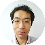
Hiratsuka, Takuya
(May 1, 2016 ~ March 31, 2020)
I was born in Kyoto Prefecture in 1974. I graduated from the Faculty of Medicine at Kyoto University in 2001, and I earned a doctorate from Kyoto University in 2008. I later worked as a pathologist at Otsu Hospital and Noe Hospital, where I gained clinical experience and encountered various illnesses. As of 2012, I became an Assistant Professor of the Pathology and Biology of Diseases in the Graduate School of Medicine at Kyoto University, and I am currently a Senior Lecturer in the Department of Drug Discovery and Medicine in the Graduate School of Medicine at Kyoto University.
At Kyoto University’s Graduate School of Medicine, I analyzed and examined the relationship between viral causes of lymphoma and an individual’s genetic background under the direction of Dr. Tsuruyama, who is now this professor. Later, I worked as a pathologist at Otsu Hospital and Noe Hospital, where I honed my skills at diagnostic pathology and when I encountered various illnesses. During that time, I became interested in the relationship between the morphological characteristics of different illnesses, protein expression and other diagnostic criteria and the mechanism of that expression, and prognosis. Starting in 2012, I worked in the Pathology and Biology of Diseases in the Graduate School of Medicine at Kyoto University. I used a transgenic mouse expressing a fluorescence resonance energy transfer (FRET) biosensor to quantitatively and qualitatively analyze protein activity in various biological phenomena and to analyze spatiotemporal regulation of that activity. I also learned how to process a variety of digital images. Using that knowledge, I want to try and create a new form of pathology here in the Department of Drug Discovery and Medicine.
A new form of pathology
AAs I mentioned earlier, I learned pathology as a pathologist. Pathology was founded in the late 19th century by the famed Thomas Hodgkin (whom Hodgkin’s disease is named after), Carl von Rokitansky, and Rudolf Virchow. Examining diseased cells under a microscope is an essential part of pathology today, but at the time it was an innovative approach to medical research. The founders of pathology laid the cornerstone of modern medical research.
So what does pathology look like today? Today, pathology is both part of basic medicine and it plays a role in clinical medicine in the area of diagnostics. Diagnostic pathology has revealed even though two cancers may be of the same type, their prognosis may differ depending on the cells that comprise those cancers. In addition, a technique known as immunostaining has been used to examine which cells correspond to a certain type of cancer depending on the proteins they express or a specific pattern of expression. Analysis of protein expression via immunostaining has led to molecularly targeted therapies and markers with which to predict the efficacy of those therapies. From the opposite perspective, pathologists already know which immunostaining results and morphological characteristics are indicative of an illness. Pathologists use protein markers in immunostaining and routine diagnosis. If pathologists could use those markers to ascertain the mechanisms responsible for certain patterns of morphology and the mechanisms responsible for certain illnesses, then pathologists may be able to identify biomarkers that can serve as therapeutic targets or aid in diagnosis.
As I mentioned earlier, pathology is a field that involves examining cells under a microscope. There are now numerous types of microscopes, such as the confocal microscope, the two-photon excitation microscope, the light sheet microscope, the stimulated emission depletion (STED) microscope (which is a type of super-resolution microscope that bypasses the diffraction limits of a light microscope and that won the Nobel Prize in Chemistry in 2014), and the photoactivated localization microscope (PALM). Microscopes are constantly improving. In addition, a hybrid optical microscope/mass spectrometer can use mass spectrometry to quantitatively indicate how much of a given substance is present in a specimen. I am convinced that use of conventional light microscopes and these new types of microscopes will open up new frontiers in pathology.
Given this reality, professor Tsuruyama and I are seeking to use mass spectrometry and a combination of optical microscopy/mass spectrometry to identify new biomarkers of various illnesses and new therapeutic targets. I also want to encourage greater use of digital technology in pathology. Pathology was merely qualitative, but I want to help develop techniques for quantification and automated analysis in pathology.
3rd Floor, Center for Anatomical, Pathological and Forensic Medical Researches, Graduate School of Medicine, Kyoto University
Yoshida Konoe-cho, Sakyo-ku, Kyoto 606-8501, Japan
TEL: +81-75-753-4427
EMAIL: hiratsuka.takuya.7v@kyoto-u.ac.jp


